What Is The Function Of The Atrioventricular Valves
Muz Play
Mar 28, 2025 · 5 min read
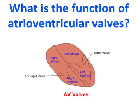
Table of Contents
What is the Function of the Atrioventricular Valves?
The human heart, a tireless engine of life, relies on a sophisticated system of valves to ensure unidirectional blood flow. Among these crucial components are the atrioventricular (AV) valves, vital structures that regulate the passage of blood between the heart's atria and ventricles. Understanding their function is key to grasping the mechanics of the cardiovascular system and appreciating the delicate balance required for efficient circulation. This comprehensive guide delves into the intricacies of AV valve function, exploring their structure, mechanism, associated pathologies, and clinical significance.
The Anatomy of Atrioventricular Valves: A Closer Look
The heart possesses two AV valves: the tricuspid valve and the mitral valve (bicuspid valve). Their names reflect their respective structures:
The Tricuspid Valve: Guardian of the Right Side
Located between the right atrium and the right ventricle, the tricuspid valve consists of three cusps (leaflets) of fibrous tissue. These cusps are attached to strong, fibrous chords called chordae tendineae, which in turn connect to papillary muscles within the ventricular wall. This intricate arrangement ensures the valve's proper closure during ventricular contraction.
Key features of the tricuspid valve:
- Three cusps: Providing a larger surface area for closure compared to the mitral valve.
- Chordae tendineae and papillary muscles: Preventing eversion (prolapse) of the cusps into the right atrium during ventricular systole.
- Lower pressure system: Compared to the left side of the heart, resulting in less stress on the valve structure.
The Mitral Valve: The Left-Sided Sentinel
The mitral valve, situated between the left atrium and the left ventricle, has two cusps. Similar to the tricuspid valve, it's anchored by chordae tendineae and papillary muscles to prevent prolapse during ventricular contraction. However, due to the higher pressure within the left ventricle, the mitral valve faces greater stress than its right-sided counterpart.
Key features of the mitral valve:
- Two cusps: Efficiently sealing the orifice between the left atrium and left ventricle.
- Robust chordae tendineae and papillary muscles: Critical for withstanding the higher pressure of the left ventricle.
- Higher pressure system: Subject to higher pressure demands, making it susceptible to certain pathologies.
The Crucial Function: Ensuring Unidirectional Blood Flow
The primary function of the AV valves is to prevent the backflow of blood from the ventricles into the atria during ventricular contraction (systole). This ensures that blood continues its journey through the circulatory system in the correct direction. Let's break down the precise mechanics:
Diastole: The Valves Open
During diastole, the relaxation phase of the cardiac cycle, the pressure within the atria exceeds that of the ventricles. This pressure difference causes the AV valves to open, allowing blood to passively flow from the atria into the ventricles. The cusps are held open by the pressure gradient, allowing for efficient ventricular filling.
Systole: The Valves Close
As the ventricles begin to contract during systole, the pressure within them rapidly increases. This pressure rise forces the AV valves to close. The chordae tendineae and papillary muscles play a critical role here, preventing the cusps from inverting (prolapsing) into the atria. The tight closure of the AV valves prevents the backflow of blood, maintaining the forward momentum of circulation.
The Role of Papillary Muscles and Chordae Tendineae
The papillary muscles and chordae tendineae are integral to the proper functioning of the AV valves. These structures work in concert to:
- Prevent prolapse: During ventricular contraction, the papillary muscles contract, tightening the chordae tendineae. This prevents the cusps from being forced upwards into the atria, ensuring a complete seal.
- Maintain valve integrity: The structural support provided by these components ensures the AV valves maintain their shape and function over time.
- Contribute to efficient blood flow: By preventing regurgitation, these elements ensure smooth and efficient blood flow through the heart.
Clinical Significance and Associated Pathologies
Malfunction of the AV valves can have significant clinical implications, leading to various cardiovascular disorders. Some common pathologies include:
Mitral Valve Prolapse (MVP)
MVP occurs when one or both cusps of the mitral valve bulge back into the left atrium during ventricular contraction. This can lead to mitral regurgitation (backflow of blood), causing symptoms such as shortness of breath, fatigue, and palpitations.
Mitral Stenosis
Mitral stenosis is a narrowing of the mitral valve orifice, restricting blood flow from the left atrium to the left ventricle. This can lead to left atrial enlargement and pulmonary hypertension. Symptoms may include shortness of breath, fatigue, and irregular heartbeat.
Tricuspid Regurgitation
Tricuspid regurgitation is the leakage of blood back into the right atrium from the right ventricle during systole. This can be caused by various factors, including damage to the tricuspid valve leaflets, chordae tendineae, or papillary muscles.
Tricuspid Stenosis
Tricuspid stenosis, though less common than mitral stenosis, involves narrowing of the tricuspid valve orifice, hindering blood flow between the right atrium and ventricle.
Atrioventricular Valve Disease Diagnosis and Treatment
Diagnosis of AV valve disease often involves a combination of physical examination, electrocardiography (ECG), echocardiography, and cardiac catheterization. Treatment options vary depending on the severity and type of valve disease and may include medication, surgical valve repair, or valve replacement.
The Significance of Understanding AV Valve Function
A comprehensive understanding of atrioventricular valve function is crucial for healthcare professionals and students alike. The intricate interplay between the valve structure, the supporting musculature, and the hemodynamic pressures within the heart dictates the efficiency of blood flow and overall cardiovascular health. Early detection and treatment of AV valve pathologies are essential for preventing serious complications and improving patient outcomes. Further research into the pathogenesis and treatment of AV valve diseases remains a crucial area of ongoing investigation in the field of cardiology. The ongoing advancements in minimally invasive surgical techniques and the development of novel biomaterials for valve replacement represent significant progress in the management of AV valve disorders. Continued efforts in this area will lead to improved therapeutic interventions and enhanced quality of life for patients affected by these conditions. The future of AV valve care holds great promise for improving cardiovascular health and extending life expectancy. In conclusion, understanding the intricacies of AV valve function is paramount for comprehending the complex mechanics of the heart and the importance of maintaining its optimal performance.
Latest Posts
Latest Posts
-
Why Is Mol The Abbreviation To Mle
Mar 31, 2025
-
What Is The Correct General Equation For Cellular Respiration
Mar 31, 2025
-
Which Element Is The Least Reactive
Mar 31, 2025
-
Which One Is Good Insulator Metals Metalloids Or Nonmetals
Mar 31, 2025
-
Why Are Covalent Compounds Not Conductive
Mar 31, 2025
Related Post
Thank you for visiting our website which covers about What Is The Function Of The Atrioventricular Valves . We hope the information provided has been useful to you. Feel free to contact us if you have any questions or need further assistance. See you next time and don't miss to bookmark.
