What Moves The Chromatids During Mitosis
Muz Play
Mar 28, 2025 · 6 min read
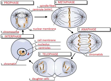
Table of Contents
What Moves the Chromatids During Mitosis? A Deep Dive into the Mechanics of Chromosome Segregation
Mitosis, the process of cell division that produces two genetically identical daughter cells, is a marvel of cellular mechanics. Central to this process is the precise and reliable segregation of duplicated chromosomes, each consisting of two sister chromatids, into the newly forming daughter cells. But what exactly moves these chromatids? This question has been a central theme in cell biology research for decades, leading to a deep understanding of the complex molecular machinery involved. This article will delve into the intricate mechanisms responsible for chromatid movement during mitosis, exploring the roles of key players like microtubules, kinetochores, motor proteins, and regulatory factors.
The Key Players: Microtubules, Kinetochores, and Motor Proteins
The movement of chromatids during mitosis is a dynamic process orchestrated by a complex interplay of several key cellular components:
1. Microtubules: The Cellular Highways
Microtubules are dynamic, hollow, cylindrical structures composed of α- and β-tubulin dimers. They form the mitotic spindle, a bipolar structure that emanates from the centrosomes at opposite poles of the cell. The spindle microtubules act as the "highways" along which chromatids are transported. There are three main types of microtubules involved:
-
Kinetochore microtubules: These microtubules attach directly to the kinetochores, specialized protein structures located at the centromeres of chromosomes. They are responsible for the precise movement of chromosomes towards the poles.
-
Polar microtubules: These microtubules extend from one pole to the other, overlapping in the middle of the cell. They help to establish and maintain the spindle's bipolar structure and generate the forces needed for pole separation.
-
Astral microtubules: These microtubules radiate outward from the centrosomes to the cell cortex. They help to position the spindle within the cell and connect the spindle to the cell membrane, ensuring proper orientation.
2. Kinetochores: The Attachment Sites
Kinetochores are intricate protein complexes assembled at the centromeres of chromosomes. They serve as the crucial interface between the chromosomes and the microtubules, mediating the attachment and movement of chromatids. The kinetochore's architecture is incredibly complex, comprising numerous proteins that interact with both microtubules and the centromeric chromatin. This sophisticated structure is essential for the accurate and error-free segregation of chromosomes. A key aspect of kinetochore function is its ability to "capture" and "stabilize" microtubules. This ensures that each chromatid is properly connected to the spindle apparatus.
3. Motor Proteins: The Engines of Movement
Motor proteins are molecular machines that convert chemical energy (ATP) into mechanical work. Several types of motor proteins play crucial roles in chromatid movement, including:
-
Kinesins: These are plus-end-directed motor proteins, meaning they move towards the plus end of microtubules (typically towards the cell periphery). Different kinesin families contribute to various aspects of mitosis, including spindle pole separation, chromosome congression, and the generation of pushing forces on chromosomes.
-
Dyneins: These are minus-end-directed motor proteins, meaning they move towards the minus end of microtubules (typically towards the centrosomes). They play a critical role in pulling chromosomes towards the spindle poles.
These motor proteins act on microtubules, generating the forces needed to move chromosomes. Their activity is highly regulated to ensure proper chromosome segregation.
The Stages of Chromatid Movement: A Step-by-Step Guide
The movement of chromatids during mitosis is not a single continuous event but rather a series of coordinated steps occurring during different phases of the cell cycle:
Prophase: Preparation for Movement
During prophase, the chromosomes condense, becoming visible under a microscope. The centrosomes duplicate and migrate to opposite poles of the cell, establishing the foundation for the mitotic spindle. Microtubules begin to polymerize, forming the spindle fibers that will eventually interact with the chromosomes.
Prometaphase: Initial Attachment and Congression
Prometaphase marks the breakdown of the nuclear envelope, allowing the spindle microtubules to interact directly with the chromosomes. Kinetochore microtubules begin to attach to the kinetochores, a process known as kinetochore capture. This process is not random; it involves a complex interplay of microtubule dynamics and motor proteins, ensuring proper bipolar attachment. Once attached, chromosomes undergo congression, a process of moving towards the metaphase plate, an imaginary plane equidistant from the two spindle poles. This movement involves a balance of pulling forces exerted by dyneins and pushing forces exerted by kinesins.
Metaphase: Alignment at the Metaphase Plate
In metaphase, chromosomes align at the metaphase plate, a critical checkpoint ensuring each chromosome is properly attached to microtubules from both spindle poles. This alignment is essential to guarantee that each daughter cell receives one copy of each chromosome. The tension generated by the bipolar attachment is sensed by the cell, providing a signal that the cell is ready to proceed to anaphase.
Anaphase: The Separation of Sister Chromatids
Anaphase is the crucial stage where sister chromatids separate and move towards opposite poles of the cell. This separation is triggered by the activation of the anaphase-promoting complex/cyclosome (APC/C), an E3 ubiquitin ligase that targets several proteins for degradation, including securin, a protein that inhibits separase. The degradation of securin allows separase, a protease, to cleave the cohesin complexes that hold the sister chromatids together. This cleavage allows the sister chromatids to separate and move towards opposite poles.
The movement of chromatids during anaphase involves two distinct phases:
-
Anaphase A: The shortening of kinetochore microtubules pulls the chromatids towards the poles. This involves the depolymerization of microtubules at the kinetochores.
-
Anaphase B: The spindle poles move further apart, contributing to the separation of chromosomes. This involves the sliding of overlapping polar microtubules, driven by motor proteins like kinesins.
Telophase and Cytokinesis: Final Stages
In telophase, chromosomes arrive at the poles, and the nuclear envelope reforms around each set of chromosomes. Chromosomes decondense, and the mitotic spindle disassembles. Cytokinesis, the division of the cytoplasm, follows, resulting in two genetically identical daughter cells.
Regulation and Error Correction: Ensuring Accurate Chromosome Segregation
The accurate segregation of chromosomes during mitosis is vital for maintaining genome stability. The process is tightly regulated by a complex network of checkpoints and error-correction mechanisms:
-
Spindle assembly checkpoint (SAC): This checkpoint monitors the attachment of kinetochores to microtubules. If errors are detected (e.g., improper attachment), the cell cycle is arrested in metaphase until the errors are corrected.
-
Aurora B kinase: This kinase plays a crucial role in the correction of erroneous kinetochore-microtubule attachments, promoting microtubule detachment and re-attachment until proper bipolar attachment is achieved.
These regulatory mechanisms are critical for ensuring the fidelity of chromosome segregation, preventing aneuploidy (abnormal chromosome numbers) and maintaining genome integrity.
Research and Future Directions
Our understanding of the molecular mechanisms driving chromatid movement during mitosis has advanced significantly, yet many questions remain. Ongoing research focuses on:
-
The detailed architecture and function of the kinetochore: Understanding the precise interactions between kinetochore proteins and microtubules is crucial for unraveling the intricacies of chromosome attachment and movement.
-
The regulation of motor protein activity: Investigating how the activity of kinesins and dyneins is coordinated to generate the necessary forces for chromosome movement is an important area of research.
-
The role of post-translational modifications in mitosis: Phosphorylation and other modifications of microtubule-associated proteins and motor proteins regulate their activity and are critical for the dynamic nature of chromosome movement.
In conclusion, the movement of chromatids during mitosis is a highly orchestrated and precisely regulated process involving the intricate interplay of microtubules, kinetochores, motor proteins, and regulatory factors. A deep understanding of these mechanisms is essential for comprehending fundamental aspects of cell biology and for developing strategies to combat diseases associated with mitotic errors. Continued research in this field will undoubtedly lead to further advancements in our understanding of this fundamental biological process.
Latest Posts
Latest Posts
-
Why Do Plants Love Water In Bio Terms
Mar 31, 2025
-
Identifying The Important Intermolecular Forces In Pure Compounds
Mar 31, 2025
-
Why Does Km Increase In Competitive Inhibition
Mar 31, 2025
-
What Is The Electron Configuration Of Beryllium
Mar 31, 2025
-
When Do You Consider Log Diterminants Similar
Mar 31, 2025
Related Post
Thank you for visiting our website which covers about What Moves The Chromatids During Mitosis . We hope the information provided has been useful to you. Feel free to contact us if you have any questions or need further assistance. See you next time and don't miss to bookmark.
